01
Boston Keratoprosthesis
Artificial corneal replacement for carefully selected eyes with a poor prognosis for conventional grafting and an ocular surface able to support a Boston KPro strategy.
↗Restoring vision beyond transplantation.
Advanced keratoprosthesis and ocular surface reconstruction for patients with severe corneal blindness in whom conventional transplantation has a poor prognosis.
Repeated graft failure, severe chemical injury, autoimmune cicatrizing disease and extreme ocular-surface damage can make conventional keratoplasty unlikely to succeed.
The objective is not to apply the same prosthesis to every eye, but to select the reconstructive strategy that best matches the ocular surface, visual potential and long-term risks of each patient.
Artificial corneal replacement for carefully selected eyes with a poor prognosis for conventional grafting and an ocular surface able to support a Boston KPro strategy.
↗A highly specialized staged procedure using autologous tooth and alveolar bone as biological support for the optical cylinder in severe end-stage ocular surface disease.
↗A biological-support alternative using autologous tibial bone in complex cases where an osteo-dental lamina cannot be used or is not the preferred option.
↗Combined reconstructive approaches integrating Boston keratoprosthesis with autologous oral mucosa for eyes with severe ocular surface compromise.
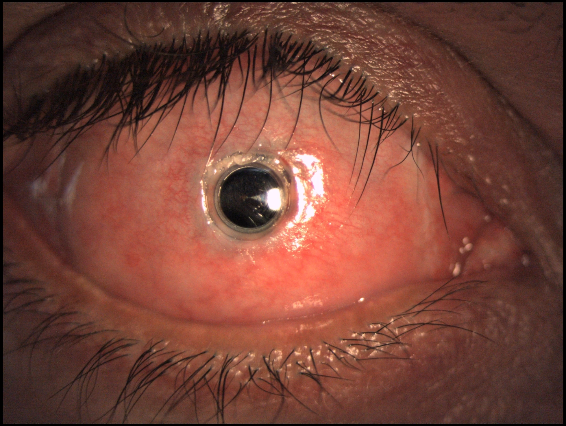
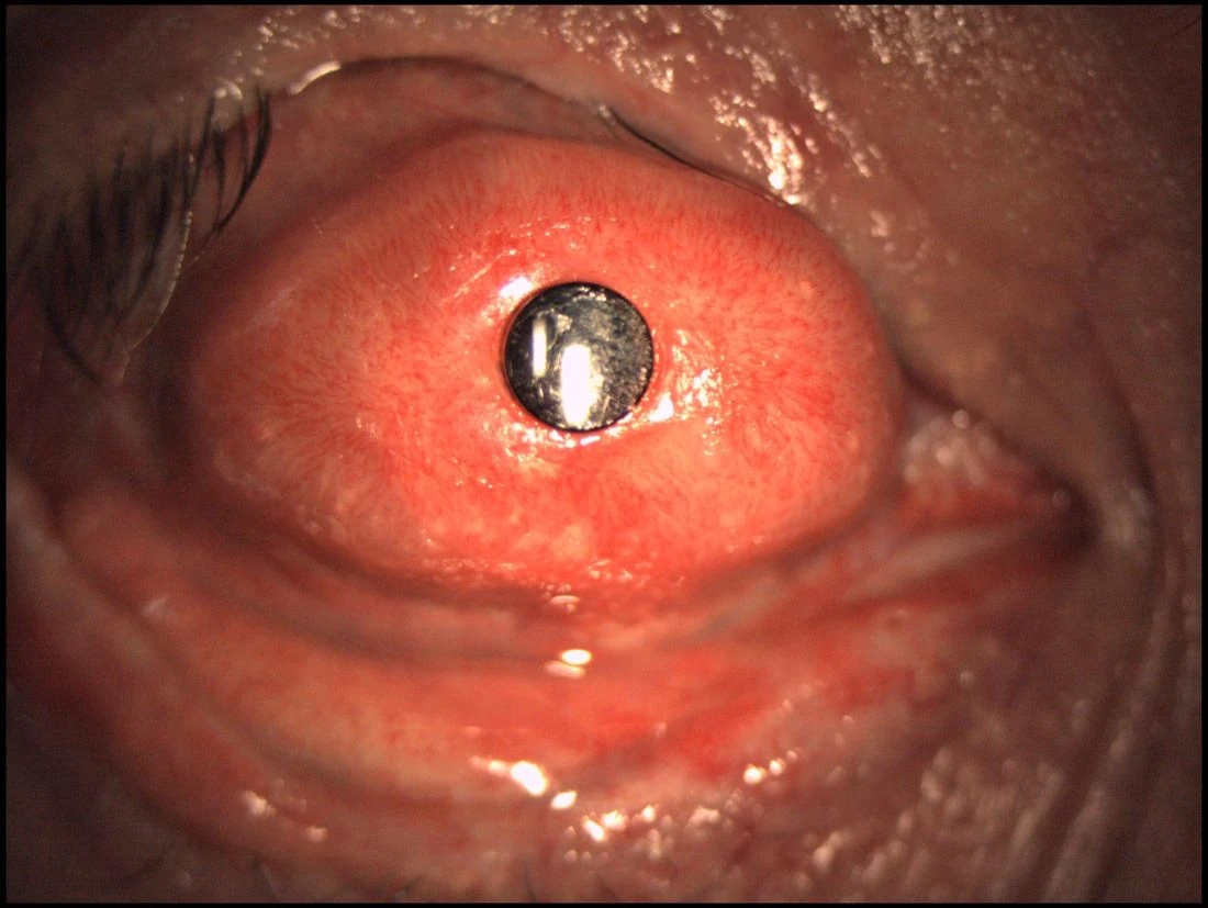
↗The Boston Type I Keratoprosthesis is one of the most established artificial cornea systems for eyes in which conventional corneal transplantation has a high risk of failure but the ocular surface remains capable of supporting a Type I approach.
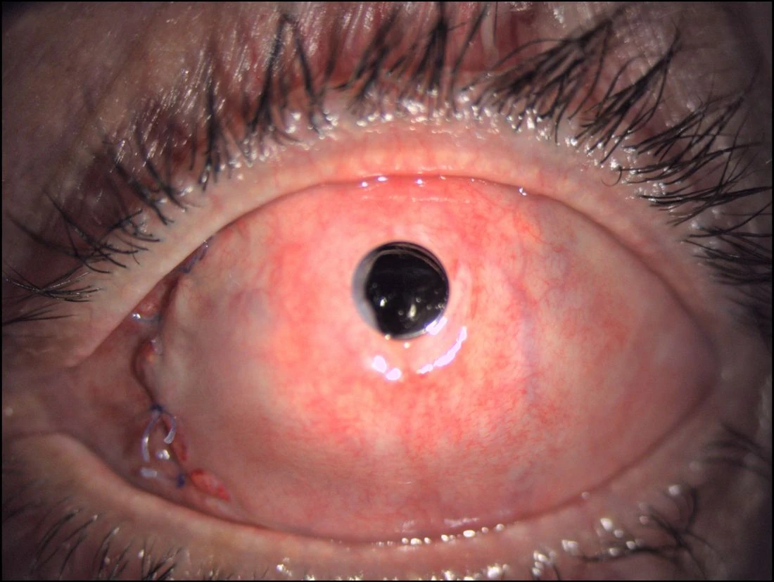
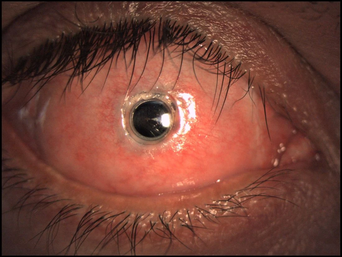
The aim of this section is to show the device in a clinical, real-world manner: not as an abstract diagram, but as a surgical solution seen at the slit lamp in actual postoperative eyes.
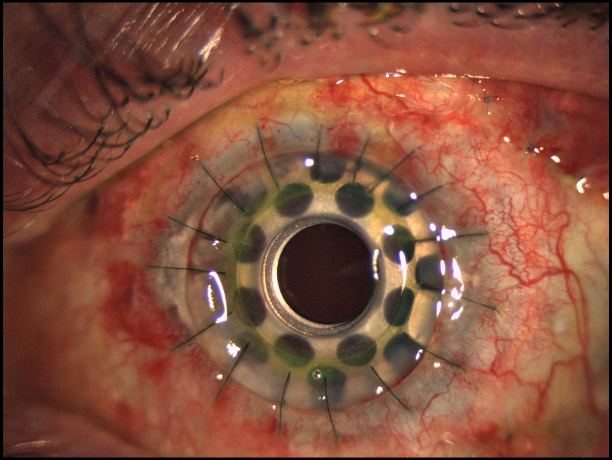
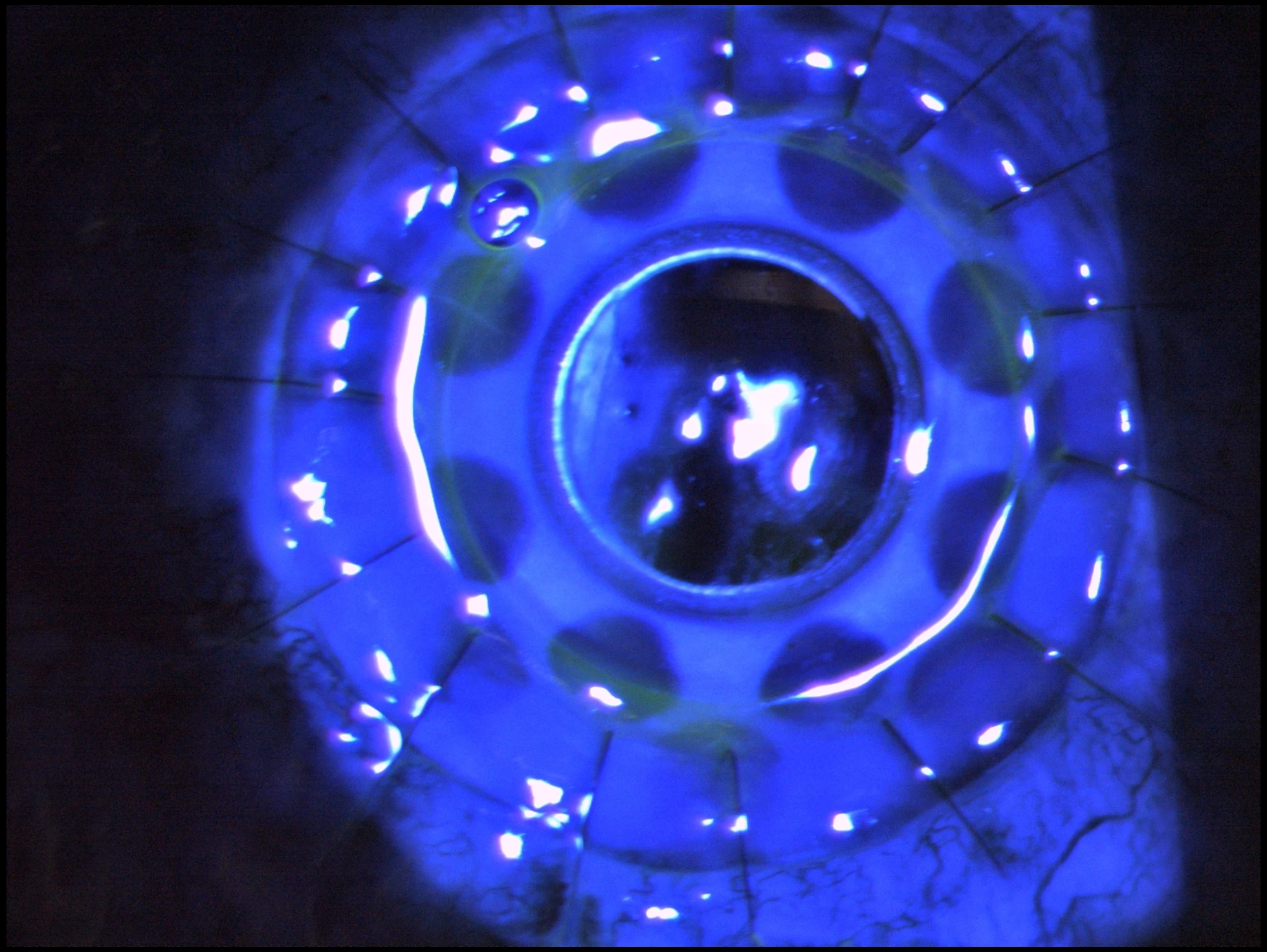
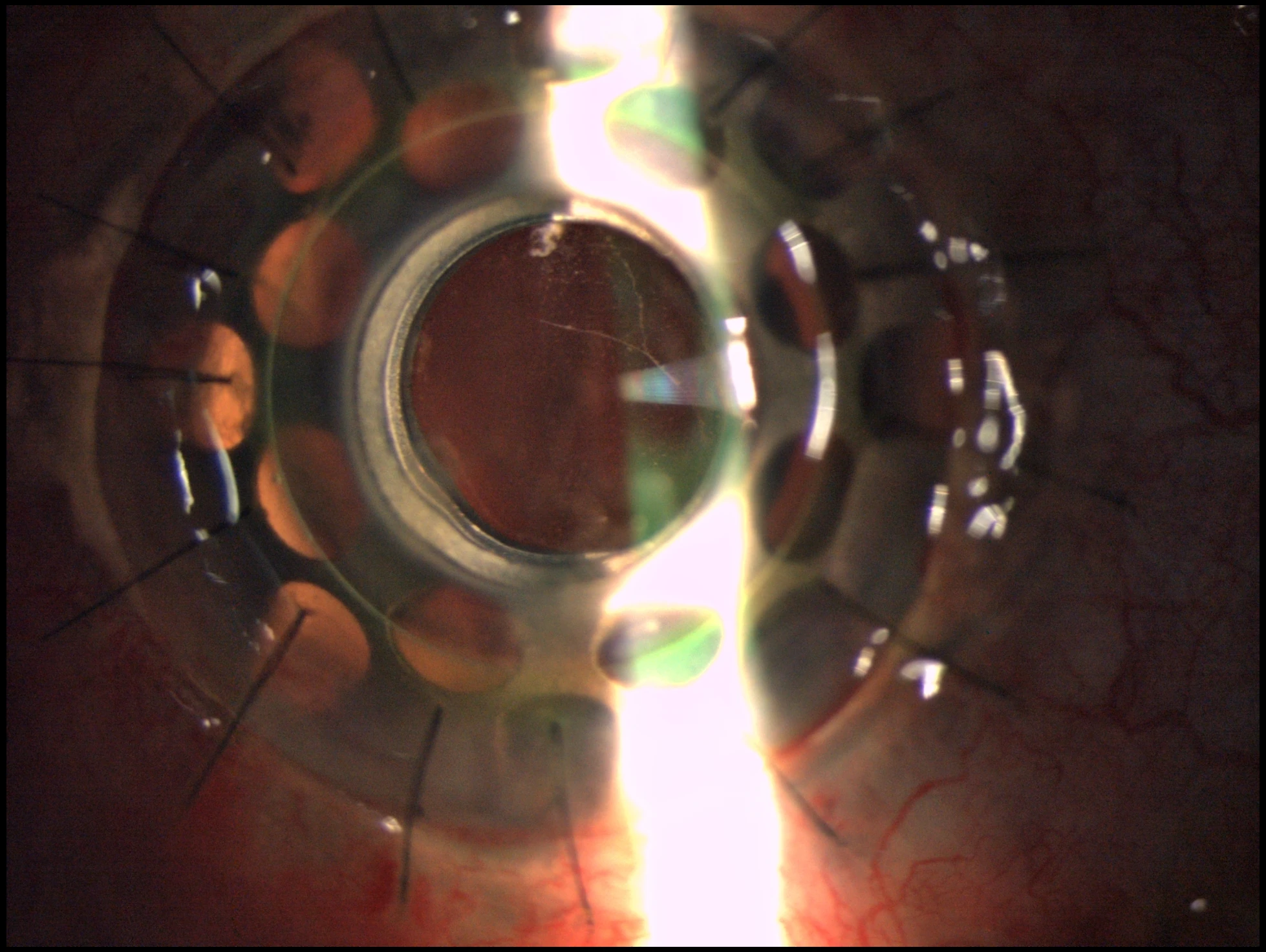
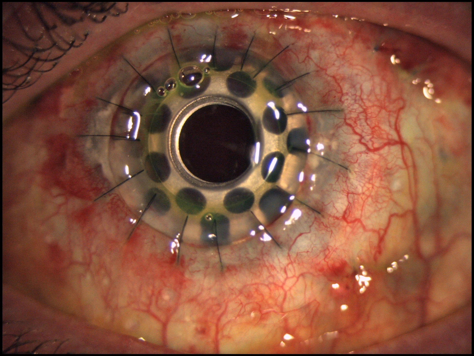
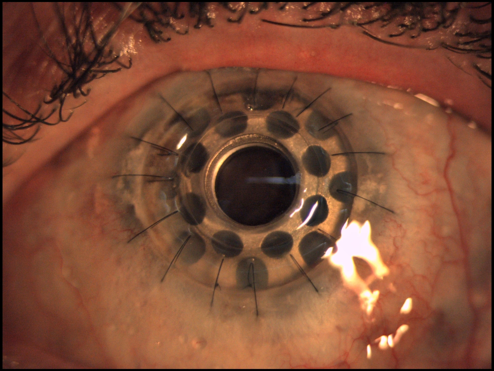
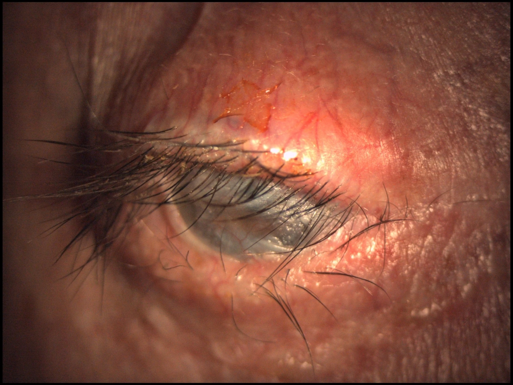
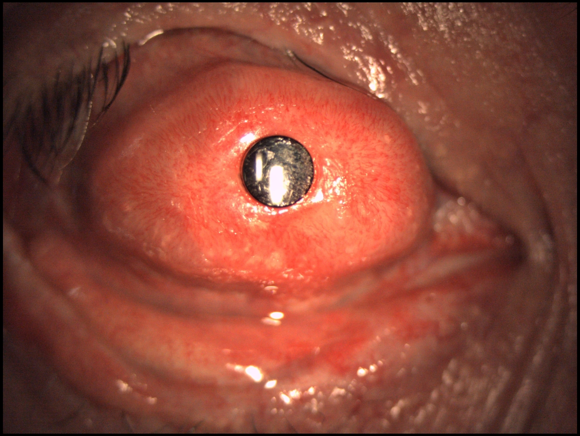
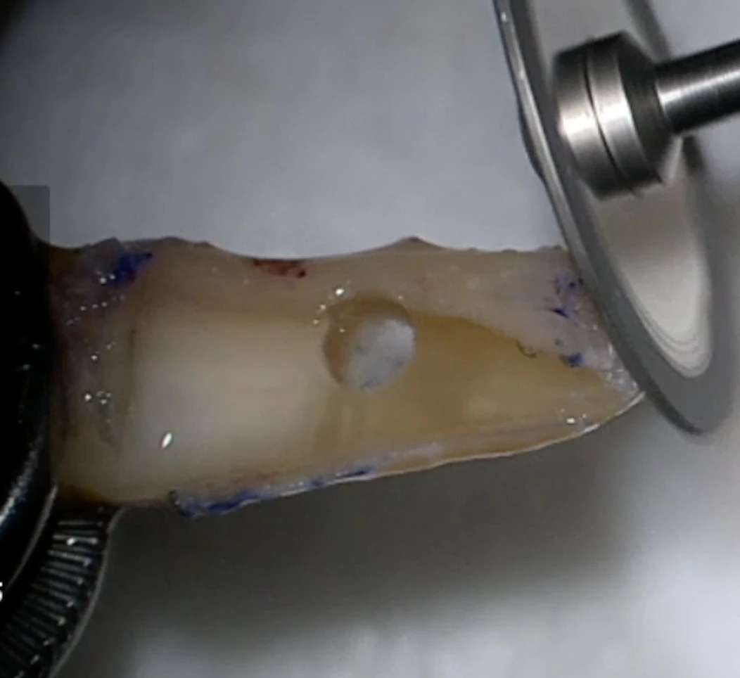
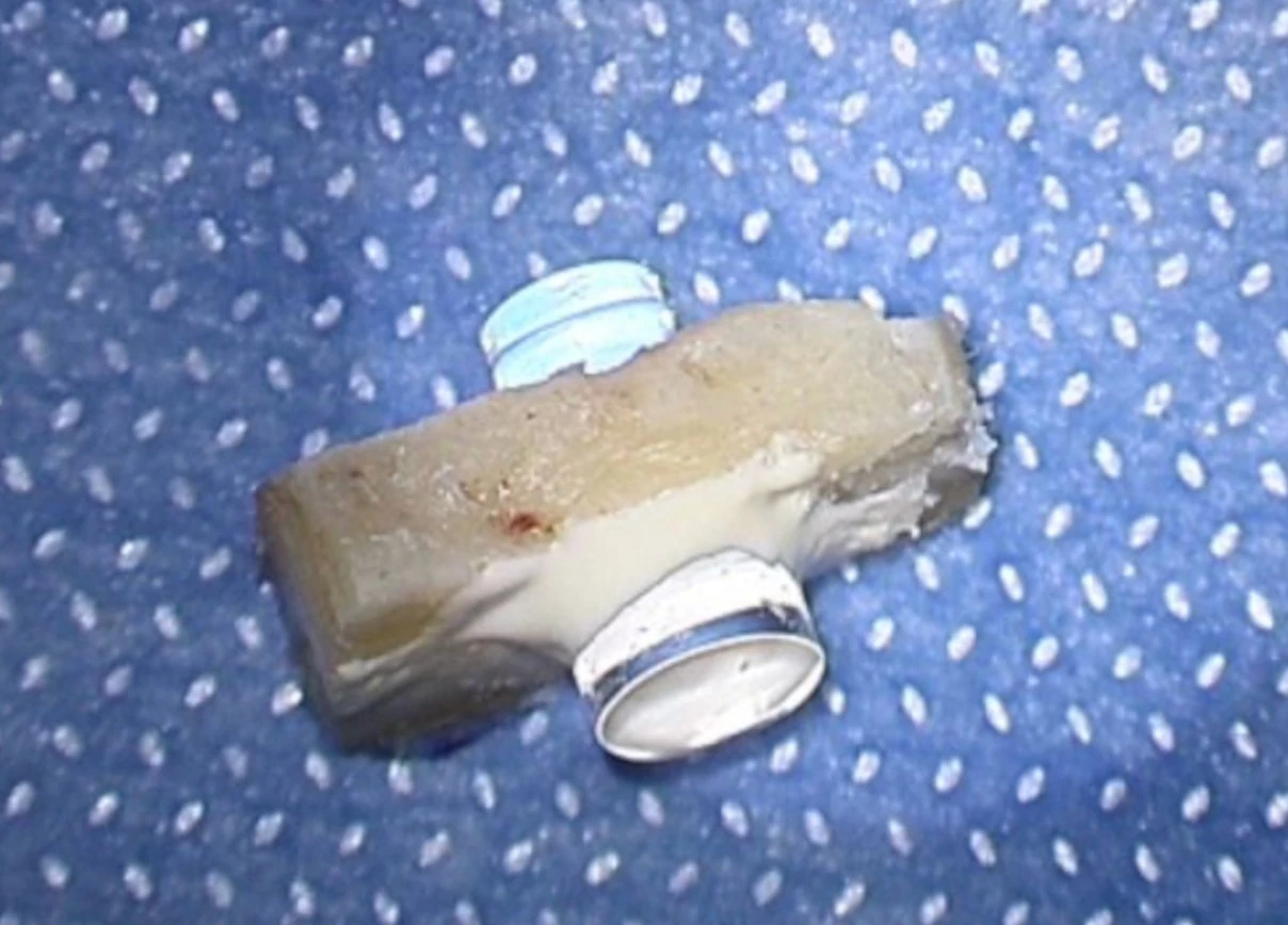
OOKP is one of the most complex and remarkable reconstructive procedures in ophthalmology. It is designed for end-stage ocular surface disease, especially in severely dry, keratinized or cicatrizing eyes in which a conventional corneal graft or even a standard Type I keratoprosthesis is unlikely to survive.
The technique uses an autologous tooth-bone lamina to support the optical cylinder and is usually combined with mucosal surface reconstruction, creating a biological environment capable of protecting the device in the most hostile ocular surfaces.
Tibial Bone Keratoprosthesis uses autologous tibial bone as the biological support for the optical cylinder. It is an alternative to osteo-odonto-keratoprosthesis in highly complex ocular-surface disease, especially when a tooth-bone lamina is not available or not ideal.
This technique belongs to the group of autologous-support keratoprostheses and is intended for very specific end-stage eyes. Its value lies in expanding the reconstructive options available when more conventional corneal procedures are no longer appropriate.
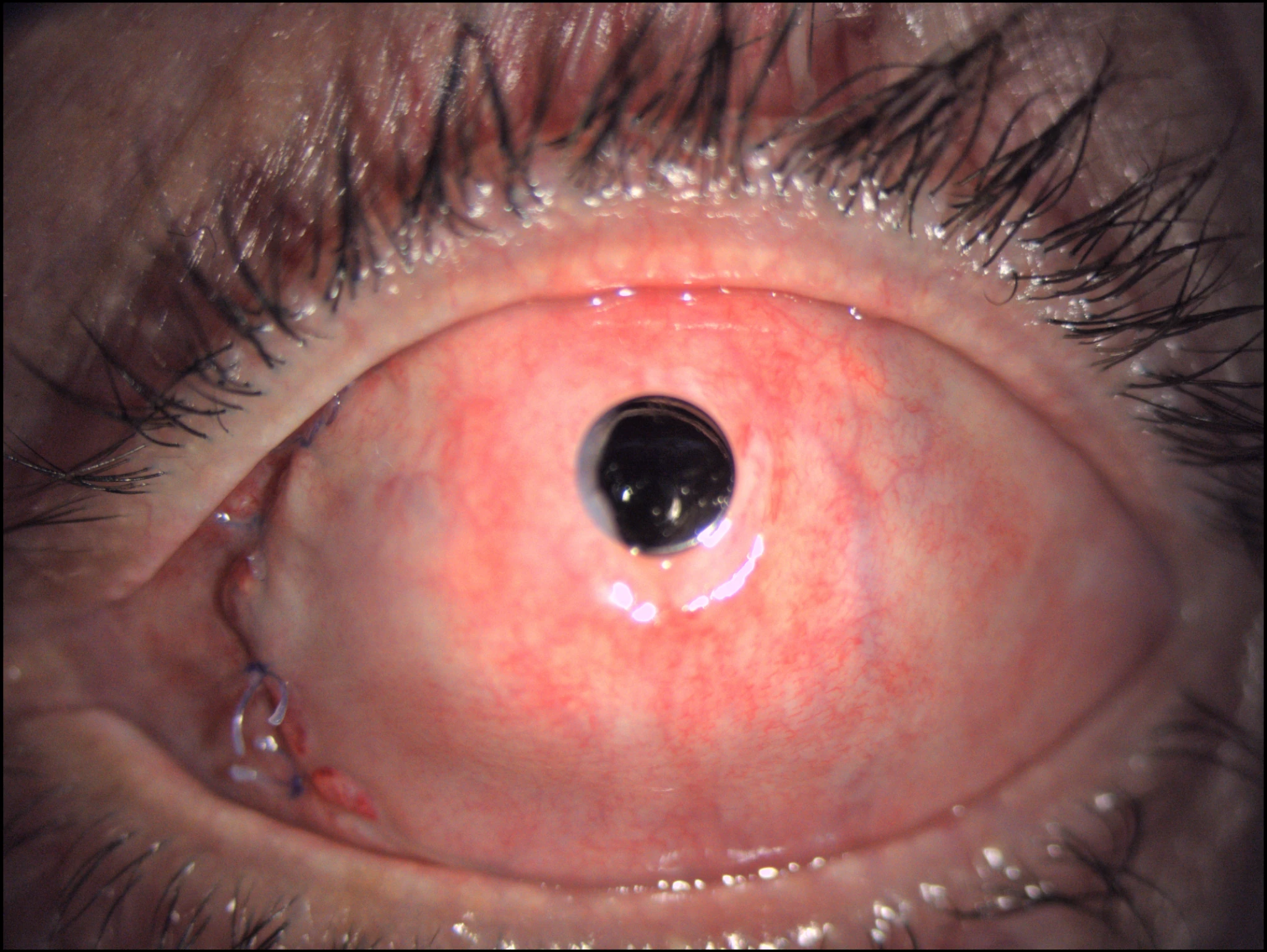
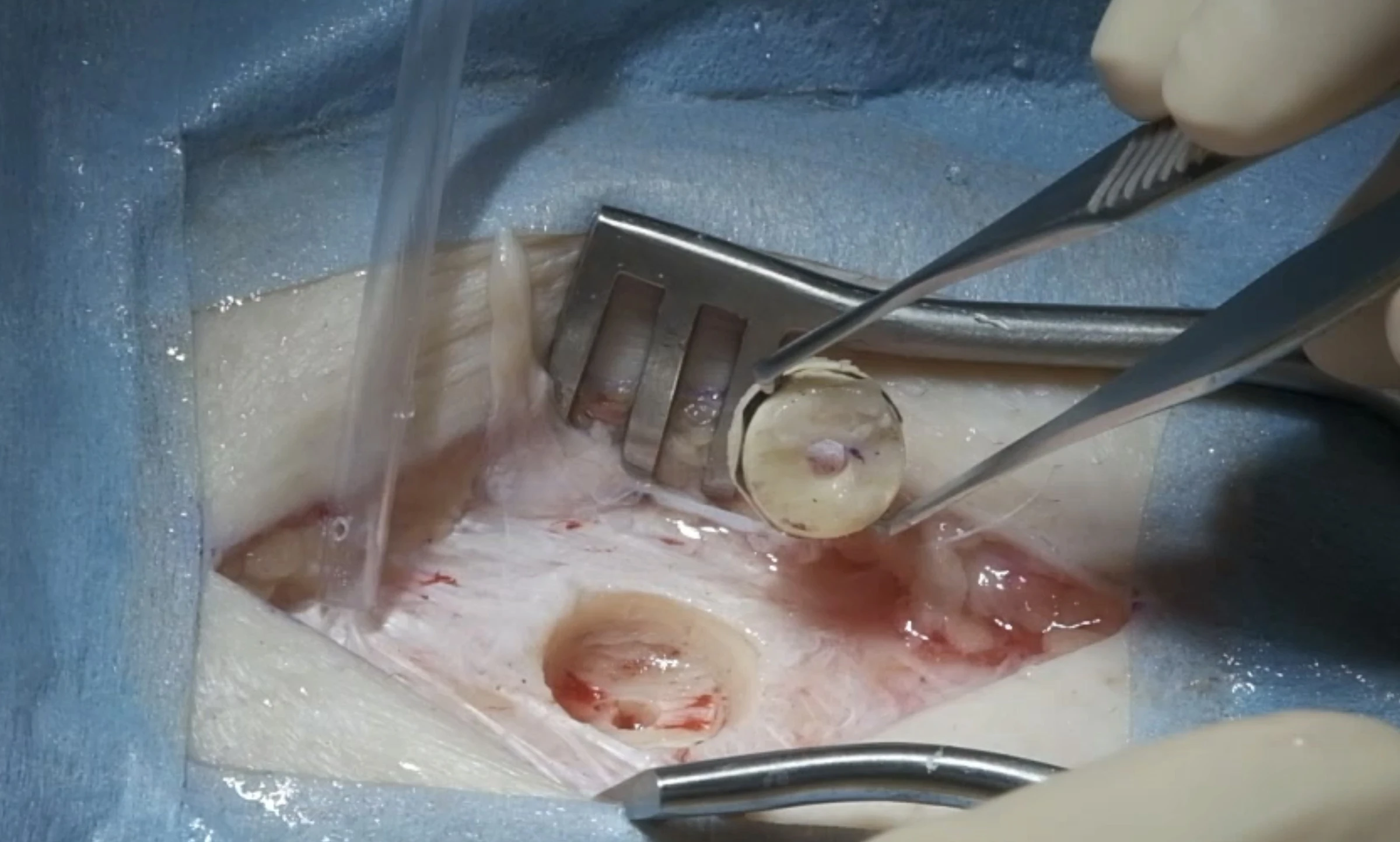
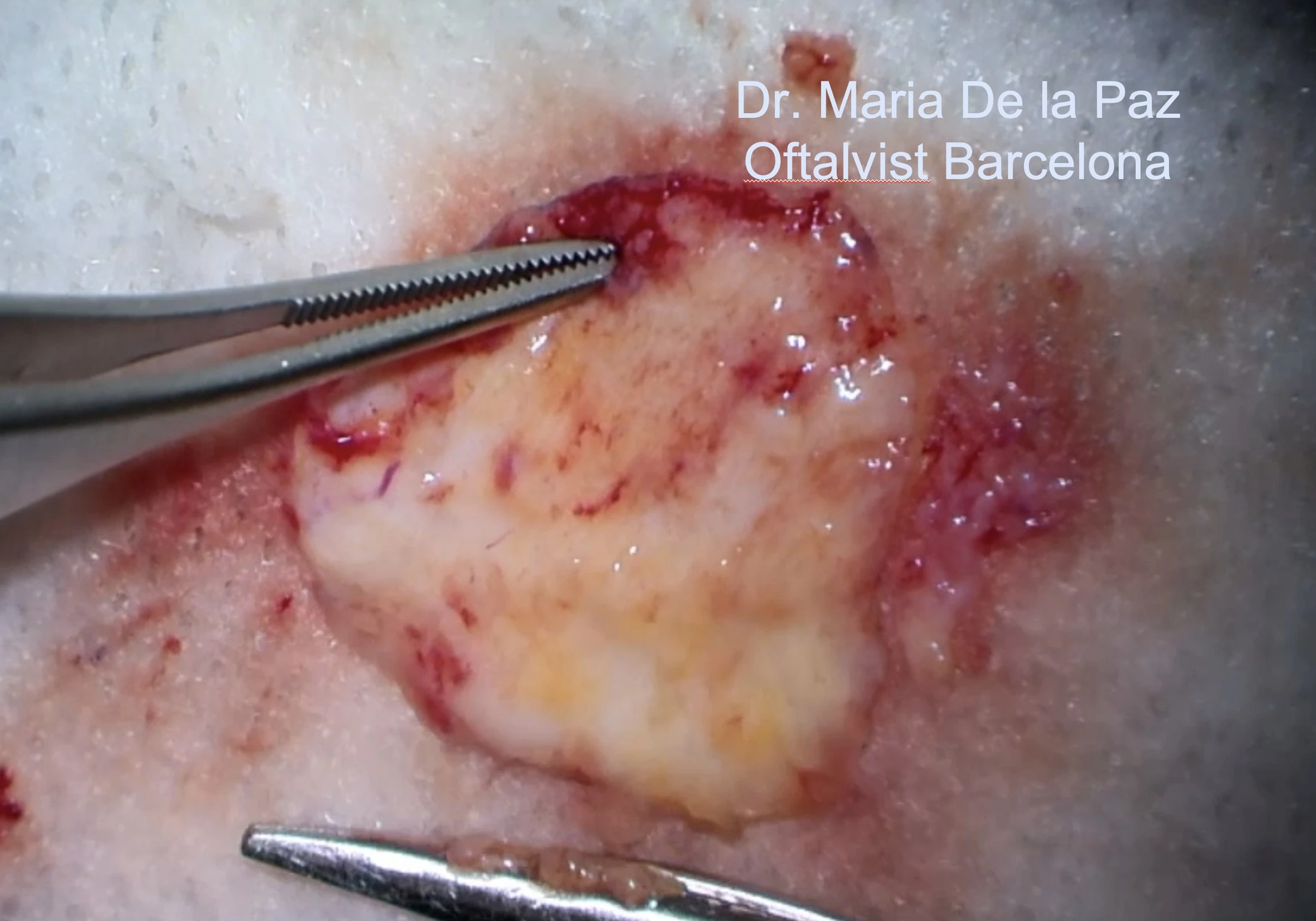
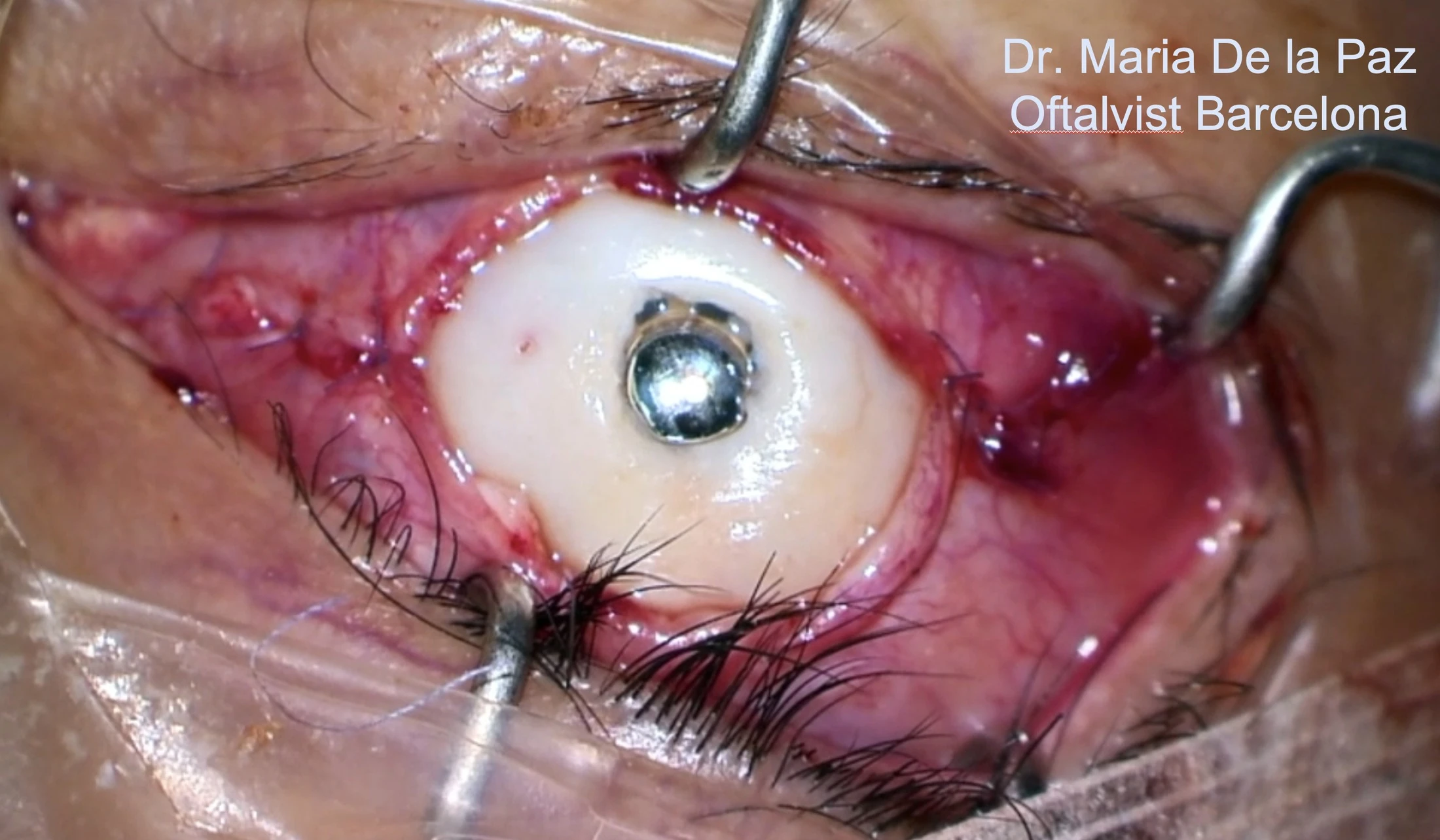
Transmucosal Boston keratoprosthesis combines the optical concept of Boston KPro with the protective and reconstructive role of autologous oral mucosa. This strategy is particularly relevant in eyes with advanced ocular-surface alteration where a standard Type I environment is not sufficient.
It represents an intermediate and highly specialized reconstructive pathway between conventional Boston KPro and the more complex autologous-support procedures such as OOKP or tibial bone keratoprosthesis.
The first decision is not “which device?”, but whether keratoprosthesis is appropriate at all. In Dr. de la Paz's clinical framework, the eye must represent end-stage ocular-surface disease with useful retinal and optic-nerve potential. The ocular surface phenotype then becomes the key determinant of the prosthetic strategy.
Functional retina and optic nerve are prerequisites before undertaking end-stage ocular-surface reconstruction.
Patient selection precedes device selection.These categories are not interchangeable. Surface wetness, blinking and keratinization, together with the underlying disease and visual potential, determine the direction of treatment.
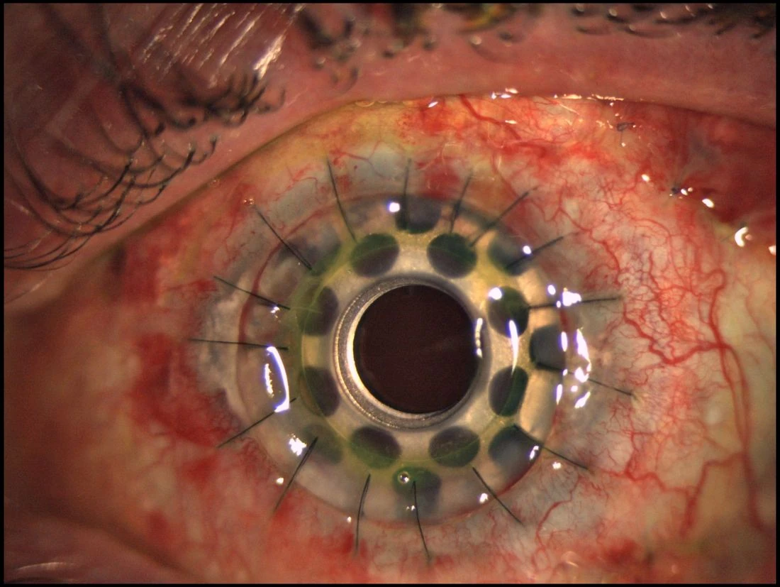
This is an educational referral framework, not an automatic surgical indication. Final choice requires a complete ocular-surface, glaucoma, retinal and systemic assessment.
A preliminary medical evaluation can be performed online. If surgery appears potentially appropriate, a complete in-person assessment can then be arranged in Barcelona.
Advanced corneal blindness requires a strategy beyond conventional transplantation.
Dr. de la Paz is an anterior segment, cornea and ocular surface surgeon based in Barcelona. Her clinical work includes limbal transplantation, ocular surface reconstruction, penetrating keratoplasty, Boston keratoprosthesis, osteo-odonto-keratoprosthesis and tibial bone keratoprosthesis.
Each surgical strategy is linked to peer-reviewed publications and long-term clinical experience. Select an article below to open its PubMed record.
Seminars in Ophthalmology.
American Journal of Ophthalmology.
Graefe's Archive for Clinical and Experimental Ophthalmology.
International surgical activity and invited lectures reinforce the site as a professional referral platform rather than a conventional clinic website.
Patients and referring ophthalmologists can request an in-person consultation in Barcelona or an online preliminary evaluation of the case. Clinical reports, slit-lamp photographs, imaging and previous surgical history can be reviewed before travel.