Advanced Keratoprosthesis & Ocular Surface Reconstruction

Which Keratoprosthesis?

Keratoprosthesis selection begins with visual potential and ocular surface phenotype. The choice is individualized according to wetness, blinking, keratinization, underlying disease and reconstructive burden.

Which Keratoprosthesis?
Clinical overview

Clinical decision framework

The first decision is not “which device?”, but whether keratoprosthesis is appropriate at all. In Dr. de la Paz's clinical framework, the eye must represent end-stage ocular-surface disease with useful retinal and optic-nerve potential. The ocular surface phenotype then becomes the key determinant of the prosthetic strategy.

Gate 1 · Is there realistic visual potential?

Functional retina and optic nerve are prerequisites before undertaking end-stage ocular-surface reconstruction.

Patient selection precedes device selection.
Wet blinking eye

Preserved surface environment

  • Complete blink
  • No significant keratinization
  • Complete lid closure
  • Inflammation under control
Primary KPro pathway → Boston KPro Type I
Dry non-blinking eye

Severely cicatrized surface

  • Severe ocular-surface fibrosis
  • Symblepharon / ankyloblepharon
  • Keratinization
  • Severe dry eye or poor lid closure
Advanced reconstructive pathway → OOKP · Tibial KPro · Boston Type II / transmucosal strategy
High riskAutoimmune disease: SJS, ocular cicatricial pemphigoid, Mooren ulcer, Sjögren syndrome.
Intermediate riskSevere chemical injury.
Lower-risk KPro groupRepeated graft failure, aniridia, silicone-oil keratopathy, LSCD, herpetic opacity and other non-autoimmune causes.
Side-by-side

Four reconstructive strategies

These categories are not interchangeable. Surface wetness, blinking and keratinization, together with the underlying disease and visual potential, determine the direction of treatment.

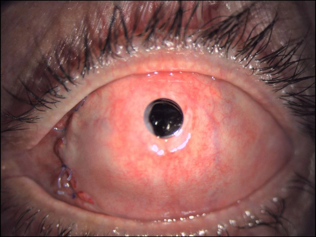
Boston KPro Type I
Wet blinking eye

Boston KPro Type I

Best surface profileComplete blink, no keratin, complete lid closure, controlled inflammation.
Main roleFirst prosthetic category considered when a wet ocular surface can support Type I KPro.
Relative burdenLess reconstructively complex than autologous-support KPro, but lifelong surveillance remains essential.
Key surveillanceGlaucoma, infection, posterior segment and device/surface complications.
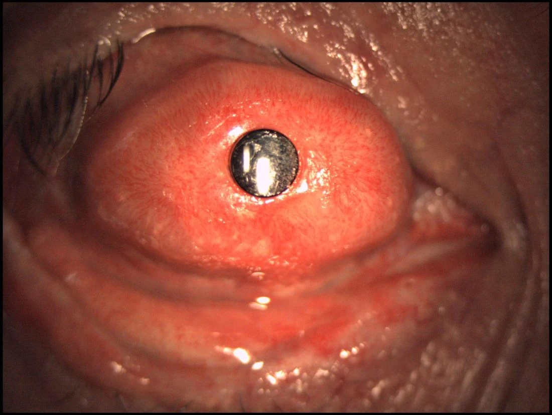
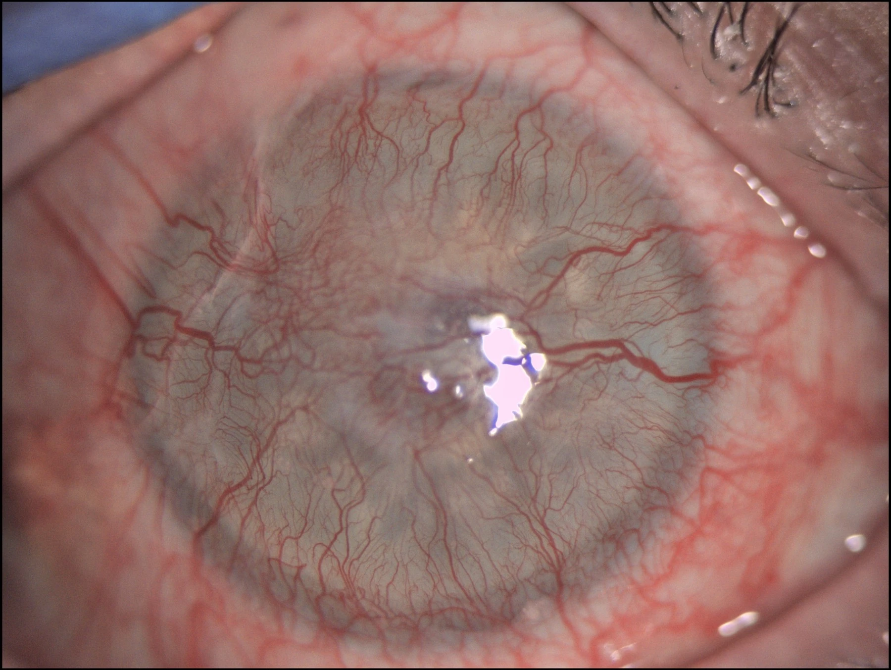
Osteo-Odonto-Keratoprosthesis
Dry non-blinking eye

OOKP

SupportAutologous tooth-bone lamina supporting an acrylic optic.
Main strengthTrue biological integration with minimal intraocular foreign material; systemic immunosuppression is not mandatory.
Trade-offsVery steep learning curve, smaller visual field, cosmetic limitations, difficult reversibility and difficult repetition.
Key surveillanceMucosal problems, extrusion, bone resorption, infection, glaucoma and posterior-segment complications.
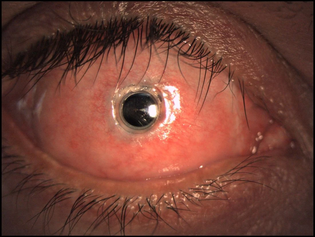
Tibial Bone Keratoprosthesis
Dry non-blinking eye

Tibial Bone KPro

SupportAutologous tibial bone supporting the optical cylinder.
Main roleBiological-support KPro in the same severe dry-eye spectrum as OOKP.
Clinical contextUseful within the autologous-support pathway when the reconstructive plan favors tibial bone rather than osteo-dental support.
Key surveillanceMucosal complications, extrusion, bone resorption, infection, glaucoma, RD/CME and vitritis.
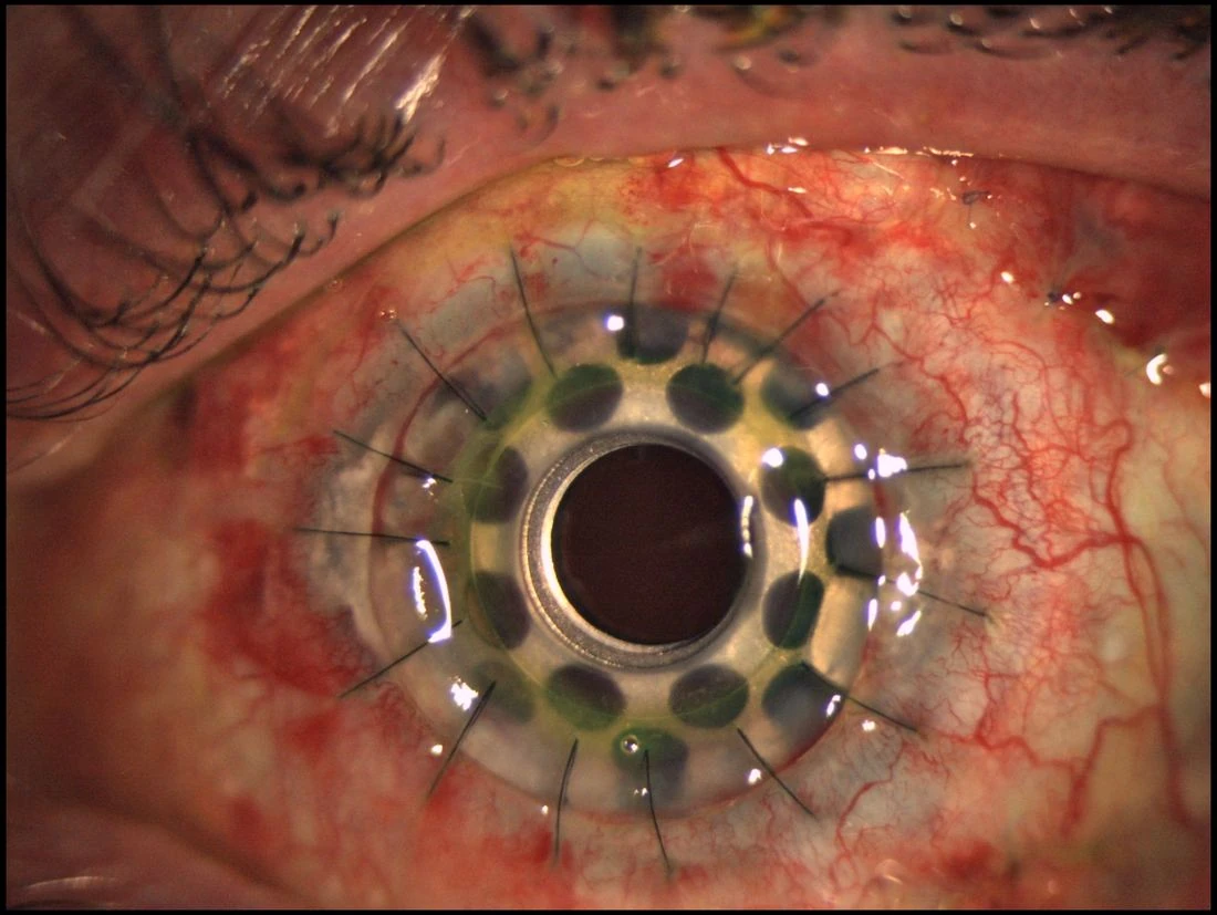
Boston KPro with buccal mucosa
Severely altered surface

Boston KPro + Buccal Mucosa

ConceptBoston optics combined with mucosal surface reconstruction.
Clinical nicheSelected severely altered ocular surfaces requiring mucosal protection rather than a standard Type I environment.
Presentation contextDr. de la Paz places Boston Type II among options for dry non-blinking eyes; her published work also includes transmucosal Boston approaches.
Key pointA hybrid reconstructive option rather than a simple extension of standard Boston Type I.
Simplified referral algorithm

From ocular surface to KPro family

01 End-stage ocular surface disease? High graft-rejection risk, repeated graft failure, LSCD, severe burns, cicatrizing disease or failure of prior surface-reconstruction procedures.
02 Retina and optic nerve functional? If visual potential is inadequate, the rationale for a high-risk KPro procedure changes fundamentally.
03 Wet blinking or dry non-blinking? Wet → Boston Type I pathway. Dry/cicatrized → OOKP, Tibial KPro or selected transmucosal / Type II Boston strategy.

This is an educational referral framework, not an automatic surgical indication. Final choice requires a complete ocular-surface, glaucoma, retinal and systemic assessment.

Medical information reviewed for this website on 24 August 2026. Final treatment selection requires an individualized ophthalmic assessment.
Evidence & innovation

Selected scientific evidence.

Each surgical strategy is linked to peer-reviewed publications and long-term clinical experience. Select an article below to open its PubMed record.

Appointments · Barcelona & Online

Request an evaluation.

Patients and referring ophthalmologists can request an in-person consultation in Barcelona or an online preliminary evaluation of the case. Clinical reports, slit-lamp photographs, imaging and previous surgical history can be reviewed before travel.

International Department
[email protected]
Oftalvist Barcelona
C/ Anglí, 40
08017 Barcelona · Spain
HLA Internacional Barcelona · 4th floor
Please indicate whether you are requesting an in-person appointment or an online case evaluation.
Online evaluation In-person visit